
Case Studies
Real clinical cases demonstrating surgical precision, innovation, and successful patient outcomes.
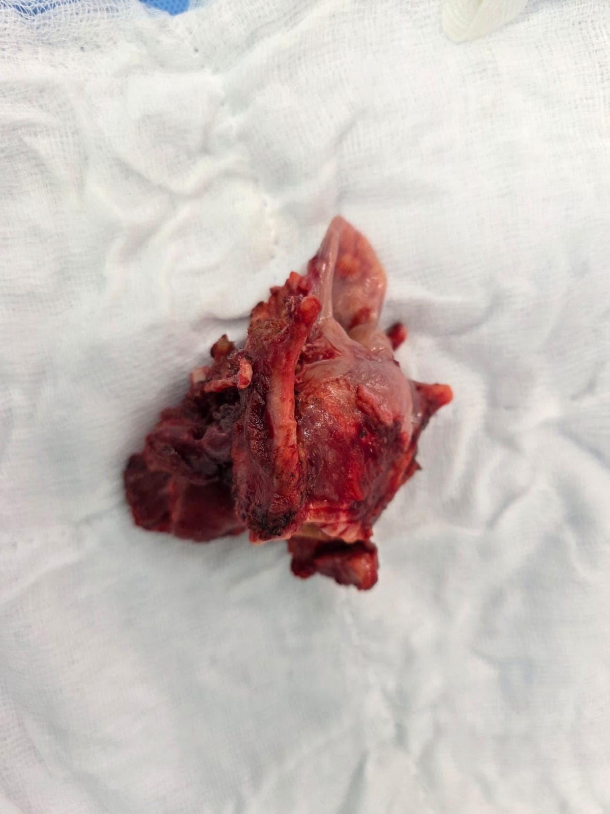
Post-Chemoradiation Failure • Laryngectomy • Pharyngeal Wall Reconstruction
The patient presented with recurrent disease involving the right pyriform fossa following failure of chemoradiation therapy. The patient underwent laryngectomy with resection of the involved pharyngeal wall to achieve complete surgical clearance. Following tumor resection, reconstruction of the pharyngeal wall was performed using a pectoralis major myocutaneous flap. The procedure was successfully completed with reconstruction of the pharyngeal defect, providing structural restoration and supporting functional recovery.
Bilateral Giant Juvenile Fibroadenoma • Multiple Breast Lesions • MED12 Exon 2 Mutation
The patient presented with a rapidly growing lump in the left breast measuring 10 × 8 cm. Eighteen months later, a second lump developed in the right breast measuring 8 × 6 cm. Both lesions were painless, firm, freely mobile masses with a bosselated surface and no associated nipple discharge or regional lymphadenopathy. Imaging and FNAC findings were suggestive of fibroadenoma. The left breast mass was surgically excised, and histopathological examination confirmed a giant juvenile cellular fibroadenoma. The patient subsequently developed another swelling in the right breast, which was also diagnosed as cellular fibroadenoma and surgically excised. Molecular analysis of the tissue identified a MED12 exon 2 mutation (c.122_139del18/p.Val41_Asn46del), providing further insight into the genetic basis of fibroadenoma. The case represents a rare presentation of bilateral giant juvenile cellular fibroadenomas with multiple recurrent lesions.
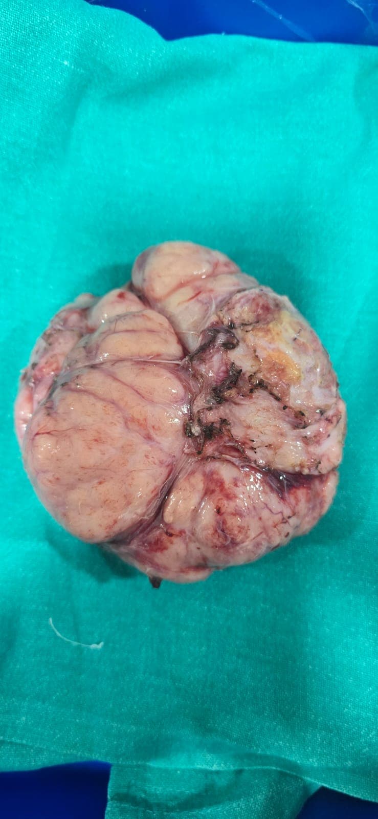
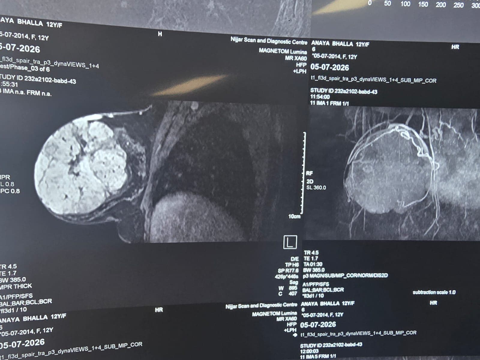
Giant Fibroadenoma • Complete Excision with Capsule Preservation
A 12-year-old girl presented with a giant breast fibroadenoma measuring 12 × 10 × 10 cm. Giant fibroadenoma is a rare benign breast lesion, particularly in young adolescent girls. The patient underwent complete surgical excision of the lesion along with its capsule through a periareolar incision. The procedure was successfully completed with careful attention to preserving the surrounding breast tissue and achieving an optimal cosmetic outcome. The patient recovered well following surgery and remains under follow-up.
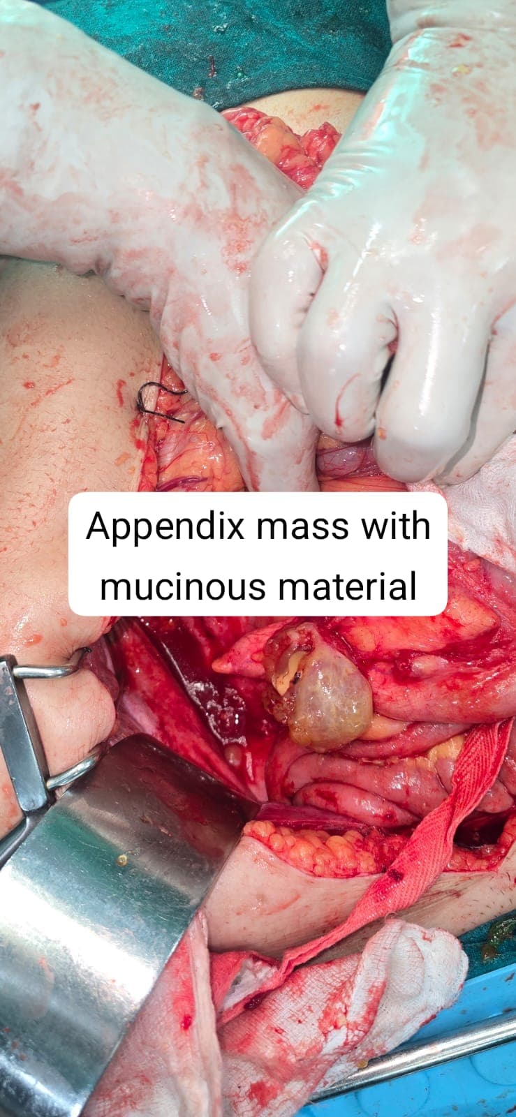
A 55-year-old woman presented with right-sided abdominal pain. CECT and PET-CT scans revealed an appendiceal mass associated with a cystic lesion and pelvic fluid. FNAC confirmed the presence of mucin, suggesting a low-grade appendiceal mucinous neoplasm. Further evaluation indicated pseudomyxoma peritonei arising from the appendiceal tumor. The patient underwent extensive cytoreductive surgery, including right hemicolectomy, omentectomy, peritonectomy, total abdominal hysterectomy, bilateral salpingo-oophorectomy, and Glisson’s capsule resection. Following complete cytoreduction, HIPEC with cisplatin was administered using the open Coliseum technique. The procedure was completed successfully, and the patient had an uneventful recovery. She was discharged in stable condition and remains under follow-up.
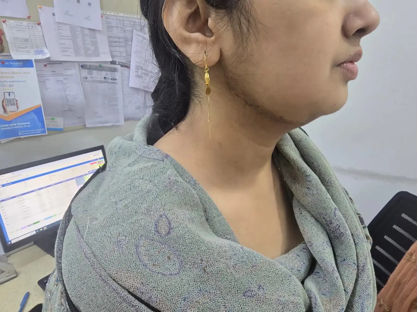
Mandible Reconstruction • Free Fibula Flap
A 32-year-old female presented with recurrent ameloblastoma after undergoing curettage five years earlier. Due to the aggressive recurrence, a segmental mandibulectomy was planned to achieve complete tumor clearance. The jaw was reconstructed using a free fibula flap to restore facial contour, speech, and chewing function. The procedure achieved good oncological safety along with functional and aesthetic rehabilitation.
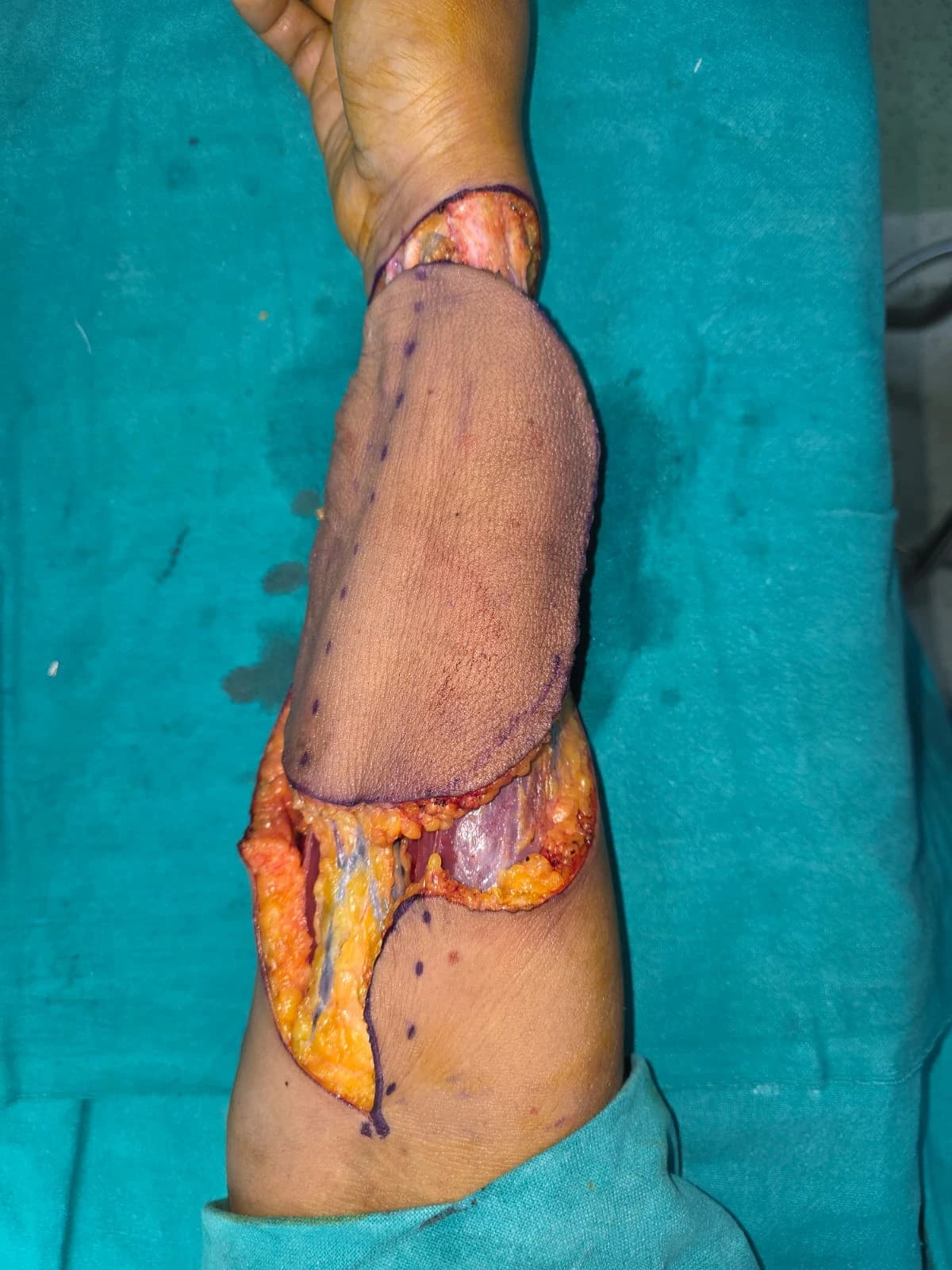
Uncommon Site • Non-Metastatic
Intermediate-grade pleomorphic soft tissue sarcoma at an uncommon elbow location. PET-CT confirmed a non-metastatic disease, with the tumor involving the overlying skin while sparing the bone. A wide local excision was performed with clear margins confirmed on frozen section. Both radial and ulnar nerves were carefully preserved to maintain limb function. The post-excision defect was reconstructed using a pedicled radial artery forearm flap, ensuring good coverage and functional recovery.
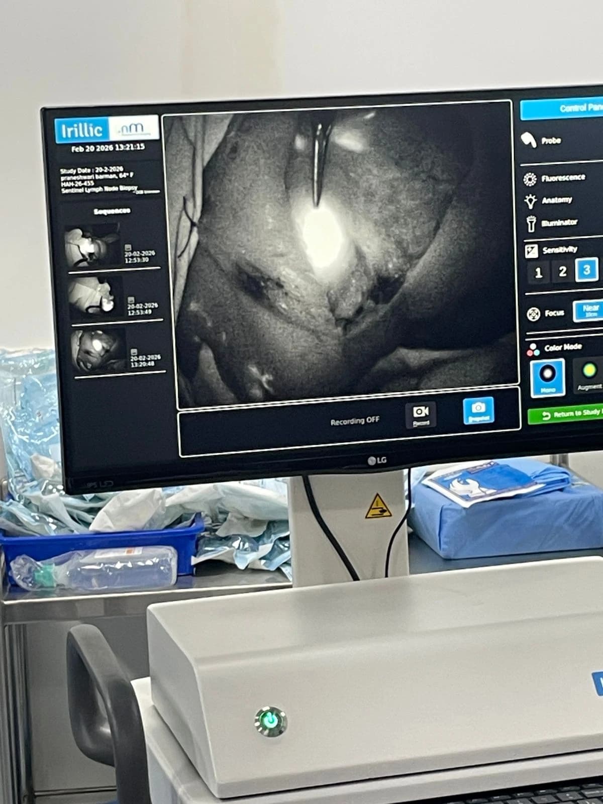
Breast Cancer • Fluorescence Guided Surgery
Sentinel lymph node biopsy was performed in a patient with early breast cancer using a dual-tracer technique with Indocyanine Green (ICG) and Methylene Blue dye. ICG fluorescence imaging helped in real-time mapping of lymphatic drainage and accurate identification of sentinel lymph nodes. The mono fluorescence mode highlights lymphatic channels clearly, while the pseudo-color mode enhances visual contrast for precise localization during surgery. This technique allows targeted removal of only the first draining lymph nodes, helping avoid unnecessary axillary lymph node dissection. The approach reduces surgical trauma, lowers the risk of arm swelling (lymphedema), and improves postoperative recovery while maintaining oncological safety.